
What the Gastrocnemius Muscle Does and Why It Matters
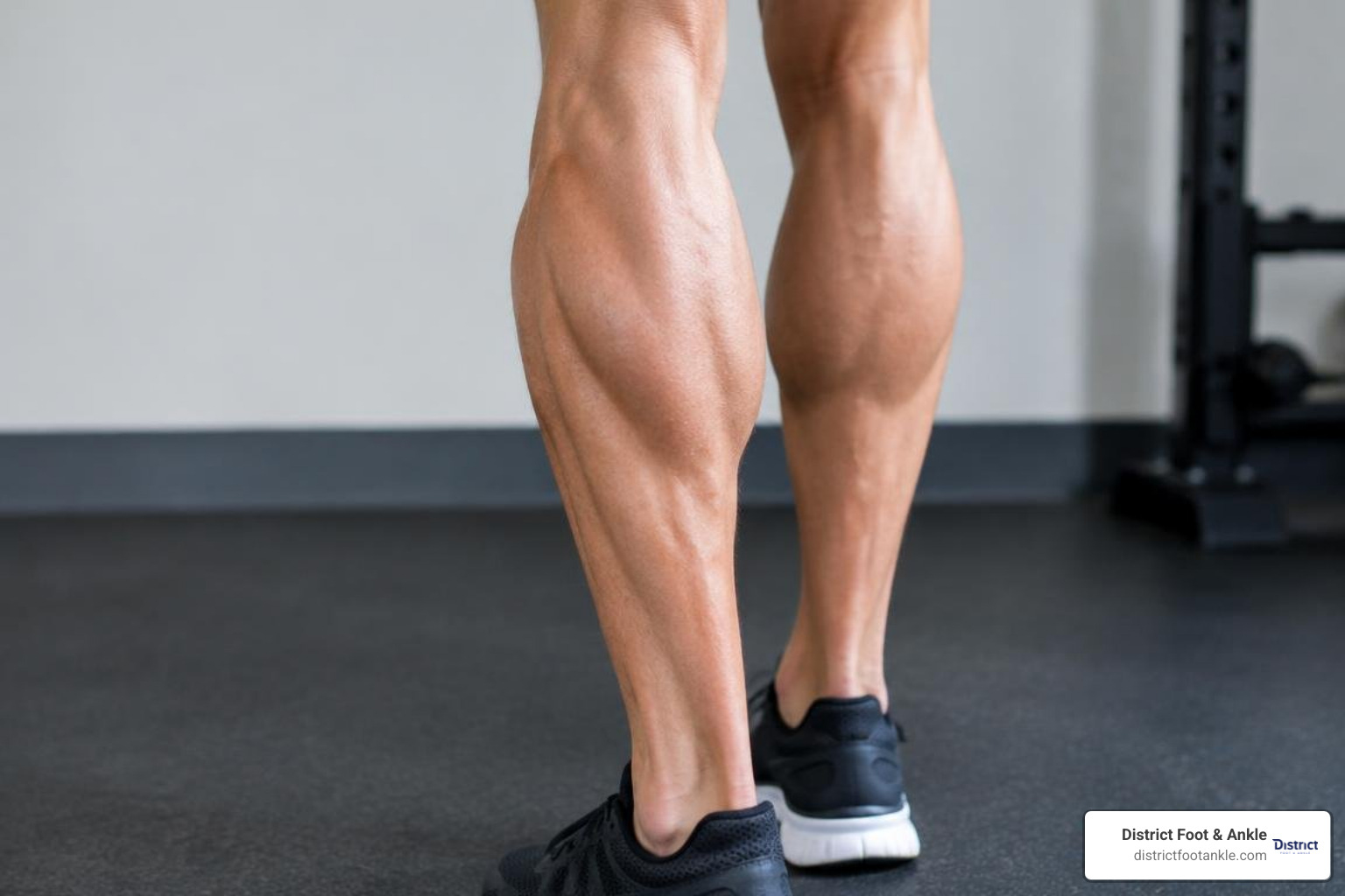
The gastrocnemius muscle is the large, visible calf muscle on the back of your lower leg. It crosses both the knee and ankle, helping you bend the knee and point the foot downward during walking, climbing stairs, running, jumping, and pushing off on a bike pedal.
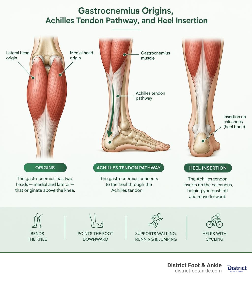
It has two heads, called the medial and lateral heads. Together with the deeper soleus muscle, it connects to the heel through the Achilles tendon. Because it handles fast, forceful movement, a tight or strained gastrocnemius can make everyday activities and sports painful.
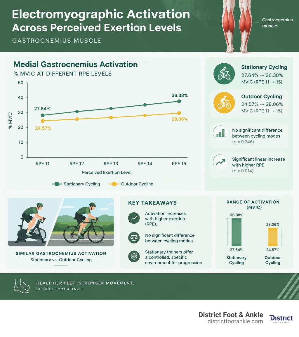
For cyclists, calf activity rises as perceived effort rises. Research comparing stationary and outdoor cycling found that medial gastrocnemius activation increased with higher effort, while activation did not differ significantly between the two riding settings. That is useful context for anyone rebuilding calf strength after pain or injury.
I am Lonny Nodelman, DPM, FACFAS, a board-certified podiatrist and active runner and cyclist who treats foot, ankle, Achilles, and gastrocnemius muscle problems. In the sections ahead, I will explain how this key calf muscle works, why it gets injured, and what can help you return to comfortable movement.
Structural Anatomy and Attachments of the Gastrocnemius Muscle
The gastrocnemius is the most superficial muscle located within the posterior compartment of the lower leg. Etymologically derived from the Greek words gaster (belly) and kneme (leg), it literally translates to the “belly of the leg”—an apt description for the distinct, twin-bulge contour it gives the human calf.
As a classic biarticular (two-joint) muscle, it spans both the knee (tibiofemoral) joint and the ankle (talocrural) joint. It comprises two anatomically distinct muscular bellies:
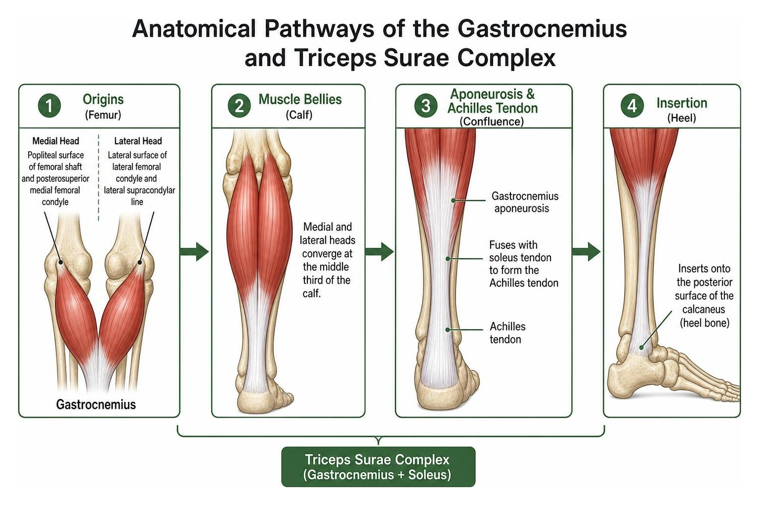
- The Medial Head: Originates proximally from the popliteal surface of the femoral shaft and the posterosuperior aspect of the medial femoral condyle. It is generally broader, extends slightly lower down the leg, and generates more sheer power than its counterpart.
- The Lateral Head: Originates from the lateral surface of the lateral femoral condyle as well as the adjacent lateral supracondylar line.
These two bellies converge inferiorly at the middle third of the calf, merging into a broad, membranous aponeurosis. This aponeurosis fuses seamlessly with the anteriorly situated tendon of the soleus muscle to form the calcaneal tendon (Achilles tendon)—the thickest, strongest tendon in the human body. The Achilles tendon subsequently inserts securely into the posterior surface of the calcaneus (heel bone). For deeper anatomical insights into its muscular architecture, see this detailed anatomy of the gastrocnemius (NBK532946).
Neurovascular Supply
The neurovascular architecture supporting the gastrocnemius is vital for both athletic performance and surgical planning:
- Innervation: The muscle receives motor innervation via the tibial nerve (spinal nerve roots S1 and S2), a terminal branch of the sciatic nerve. The tibial nerve travels down the midline of the popliteal fossa before dispatching individual muscular branches to both the medial and lateral bellies.
- Vascularization: Arterial supply is derived from the sural arteries, which branch directly off the popliteal artery within the popliteal fossa. Venous drainage empties into the popliteal vein and the small saphenous vein.
Beyond its isolated attachments, the gastrocnemius forms a continuous myofascial kinetic chain linking the plantar fascia of the foot to the hamstrings and lumbosacral fascia. Tension or tightness anywhere along this posterior line can disrupt biomechanics from the lower back down to the toes.
Variations in Gastrocnemius Muscle Morphology
Human anatomy frequently displays intriguing deviations from textbook models. The posterior compartment of the calf features several recognized morphological variants:
- The Fabella: This small sesamoid bone is embedded within the proximal tendon of the lateral head of the gastrocnemius. Found in approximately 10% to 39% of the population, the fabella can occasionally become a source of localized posterolateral knee pain (fabella syndrome) or irritate the adjacent common peroneal nerve.
- Gastrocnemius Tertius (Accessory Third Head): Present in roughly 10% of individuals, this extra muscle belly typically arises from the popliteal space or distal femur and merges with the medial or lateral head. While often asymptomatic, a bulky accessory head can narrow the popliteal fossa and contribute to Popliteal Artery Entrapment Syndrome (PAES), leading to exercise-induced calf claudication and vascular compromise in young athletes.
Histological Profile and Fiber Composition
The functional capabilities of skeletal muscle are heavily dictated by its underlying histological fiber composition. When we examine the triceps surae group under a microscope, we find a stark, intentional physiological divergence:
- Gastrocnemius (Fast-Twitch Dominant): Composed primarily of Type II (fast-twitch) glycolytic fibers (around 50% to 60%). These fibers contract rapidly and generate immense anaerobic force, making the muscle ideally tailored for explosive propulsion—such as sprinting, vertical jumping, and abrupt accelerations. However, this power comes at the cost of rapid metabolic fatigue and increased vulnerability to acute strain injuries.
- Soleus (Slow-Twitch Dominant): In contrast, the deeper soleus consists predominantly (often 70% to 85% or more) of Type I (slow-twitch) oxidative fibers. It is built for postural endurance, tonic stabilization against gravity, and sustained aerobic locomotion over long distances.
Biomechanical Functions: Walking, Running, and Athletic Propulsion
The gastrocnemius is an indispensable mechanical engine during dynamic movement. By traversing both the knee and ankle, it carries out multi-joint actions depending on whether the foot is planted (closed kinetic chain) or airborne (open kinetic chain):
- Plantarflexion at the Talocrural Joint: It points the foot downward, driving the body upward and forward.
- Assisted Knee Flexion: It acts as a secondary flexor of the knee joint, working synergistically with the hamstrings.
- Knee Joint Stabilization: During the early stance phase of the gait cycle, eccentric contraction of the gastrocnemius resists anterior translation of the tibia over the foot, providing dynamic structural support to the anterior cruciate ligament (ACL).
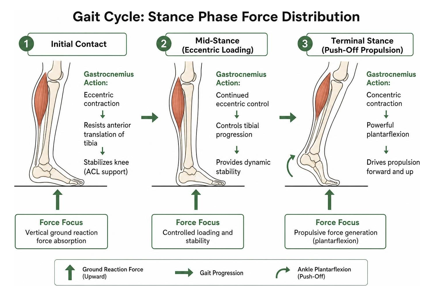
During running and sprinting, the gastrocnemius acts like a powerful biological spring. Utilizing the stretch-shortening cycle (SSC), the muscle-tendon unit rapidly absorbs ground reaction force upon footstrike and instantly releases stored elastic strain energy during push-off, minimizing metabolic energy cost while maximizing speed.
| Anatomical / Functional Characteristic | Gastrocnemius Muscle | Soleus Muscle |
|---|---|---|
| Joint Articulation | Biarticular (crosses knee and ankle) | Monoarticular (crosses ankle only) |
| Dominant Fiber Type | Type II Fast-Twitch (Explosive force) | Type I Slow-Twitch (Endurance / Posture) |
| Primary Biomechanical Role | Sprinting, jumping, dynamic acceleration | Standing posture, steady-state walking |
| Metabolic Profile | Glycolytic / Rapid fatigue | Highly oxidative / Fatigue-resistant |
| Knee Angle for Optimal Isolation | Full Knee Extension (180°) | Knee Flexion (~90°) |
| Common Clinical Pathologies | Acute muscle strains (“Tennis Leg”) | Chronic postural tightness, overuse fatigue |
Gastrocnemius Muscle Activation in Stationary vs. Outdoor Cycling
Cyclists, triathletes, and rehabilitation specialists often debate whether indoor stationary trainers recruit lower extremity musculature in the exact same manner as outdoor road cycling.
Recent electromyographic (EMG) studies have provided objective clarity on how the medial gastrocnemius behaves across these two modalities at varying self-perceived exertion levels:
- Activation vs. Cycling Mode: Medial gastrocnemius muscle activity demonstrates no statistically significant difference between overground outdoor cycling and stationary trainer cycling (p = 0.246), regardless of the rate of perceived exertion (RPE).
- Activation Across Exertion Levels: Gastrocnemius recruitment shows a significant, linear increase as intensity/RPE increases across both modes (p = 0.014).
- MVIC Activation Metrics:
- During stationary cycling, medial gastrocnemius activation ranges from 27.64% MVIC (Maximum Voluntary Isometric Contraction) at RPE 11 up to 36.38% MVIC at RPE 15.
- During outdoor cycling, activation ranges from 24.57% MVIC at RPE 11 to 28.06% MVIC at RPE 15.
Implications for Specificity and Performance
While gastrocnemius and overall power output remain consistent between stationary and outdoor conditions at submaximal workloads, other muscular differences do emerge. For instance, the gluteus maximus displays significantly higher activation during outdoor riding due to terrain micro-variations, bike sway, and stabilization demands.
For triathletes and road racers rehabilitating calf strains or targeting neuromuscular adaptations, stationary trainers provide an exceptionally specific, controlled environment to progress gastrocnemius loading without the unpredictability of road traffic and variable gradients.
Clinical Pathology, Injuries, and Assessment Techniques

Because the gastrocnemius is a superficial, biarticular muscle packed with fast-twitch fibers, it is susceptible to acute trauma and chronic overuse conditions. When calf pain or plantar tension impairs your daily movement, finding effective approaches to foot pain relief starts with an accurate diagnosis of the underlying soft-tissue structures.
1. Medial Gastrocnemius Strain (“Tennis Leg”)
“Tennis leg” is an acute, painful rupture or strain occurring at the myotendinous junction of the medial head of the gastrocnemius. It typically happens during rapid, eccentric loading—such as pushing off with a fully extended knee and dorsiflexed ankle. Patients often describe a sudden sensation resembling a direct kick or “pop” in the back of the calf, followed immediately by severe pain, swelling, and an inability to bear weight.
Clinically, muscle strains are graded according to structural severity:
- Grade I (Mild): Micro-tears of muscle fibers with minimal loss of strength.
- Grade II (Moderate): Partial tearing of muscle fibers (up to 75%) with noticeable swelling, bruising, and gait disruption.
- Grade III (Severe): Complete disruption or full tear of the muscle belly or myotendinous junction (involving >75% of muscle mass), presenting with a palpable structural defect.
To learn more about the broader spectrum of triceps surae conditions, review the clinical pathology of calf muscle injuries.
2. Gastrocnemius Equinus Contracture
Structural or functional tightness of the gastrocnemius limits upward ankle motion (dorsiflexion). This restriction forces the foot to overpronate, placing massive downstream mechanical stress on the plantar fascia, posterior tibial tendon, and metatarsals—frequently causing chronic plantar fasciitis, Achilles tendinopathy, and metatarsalgia.
3. Baker’s Cyst (Popliteal Cyst)
Fluid accumulation within the gastrocnemius-semimembranosus bursa can produce a prominent, tender swelling in the back of the knee. If a Baker’s cyst ruptures, synovial fluid dissects down into the calf tissues, closely mimicking the clinical appearance of acute deep vein thrombosis (DVT).
Diagnostic Testing and Contracture Evaluation
Accurately isolating gastrocnemius pathology requires targeted clinical and imaging assessments:
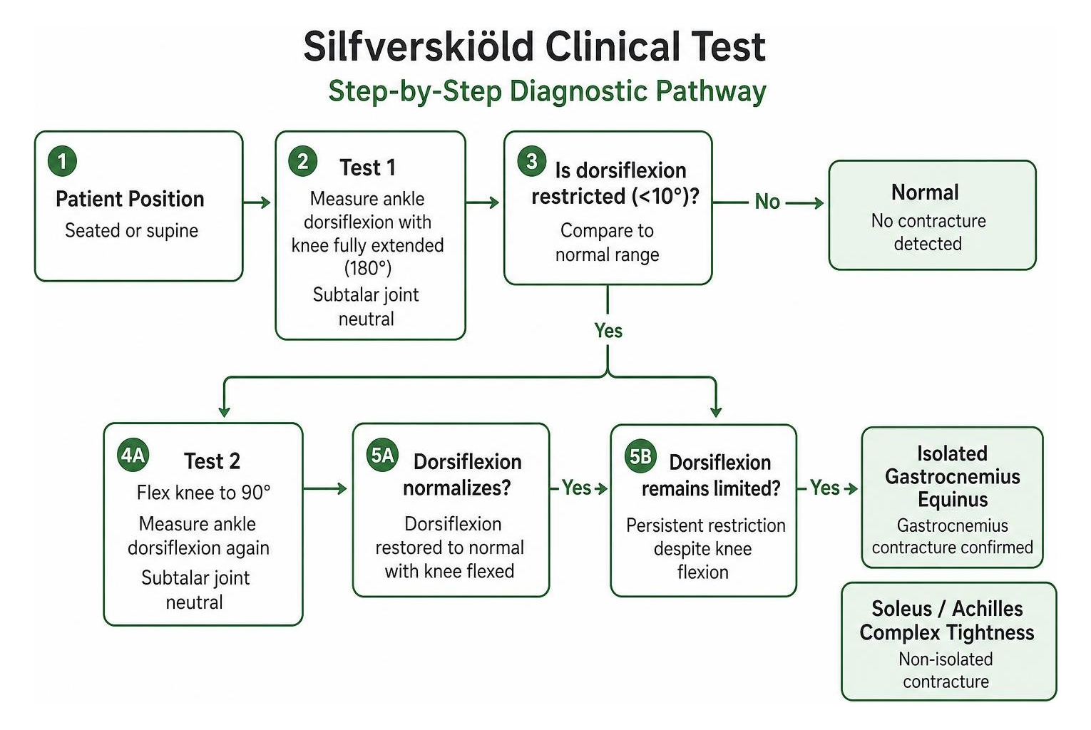
- The Silfverskiöld Test: The clinical gold standard for differentiating isolated gastrocnemius contracture from generalized Achilles/soleus tightness. With the subtalar joint held in neutral, ankle dorsiflexion is measured with the knee fully extended (180°) and then repeated with the knee flexed to 90°. If ankle dorsiflexion is restricted with the knee straight but normalizes when the knee is bent, an isolated gastrocnemius contracture is confirmed.
- The Thompson Squeeze Test: Crucial for ruling out complete Achilles tendon rupture. The clinician squeezes the calf muscle belly while the patient lies prone; failure of the foot to passively plantarflex indicates an Achilles tendon rupture (a condition misdiagnosed in up to 25% of initial presentations).
- Diagnostic Ultrasound & MRI: High-resolution point-of-care ultrasound enables real-time visualization of myotendinous fiber disruption, hematoma formation, and muscle bundle retraction.
Rehabilitation, Loading, and Strength Conditioning
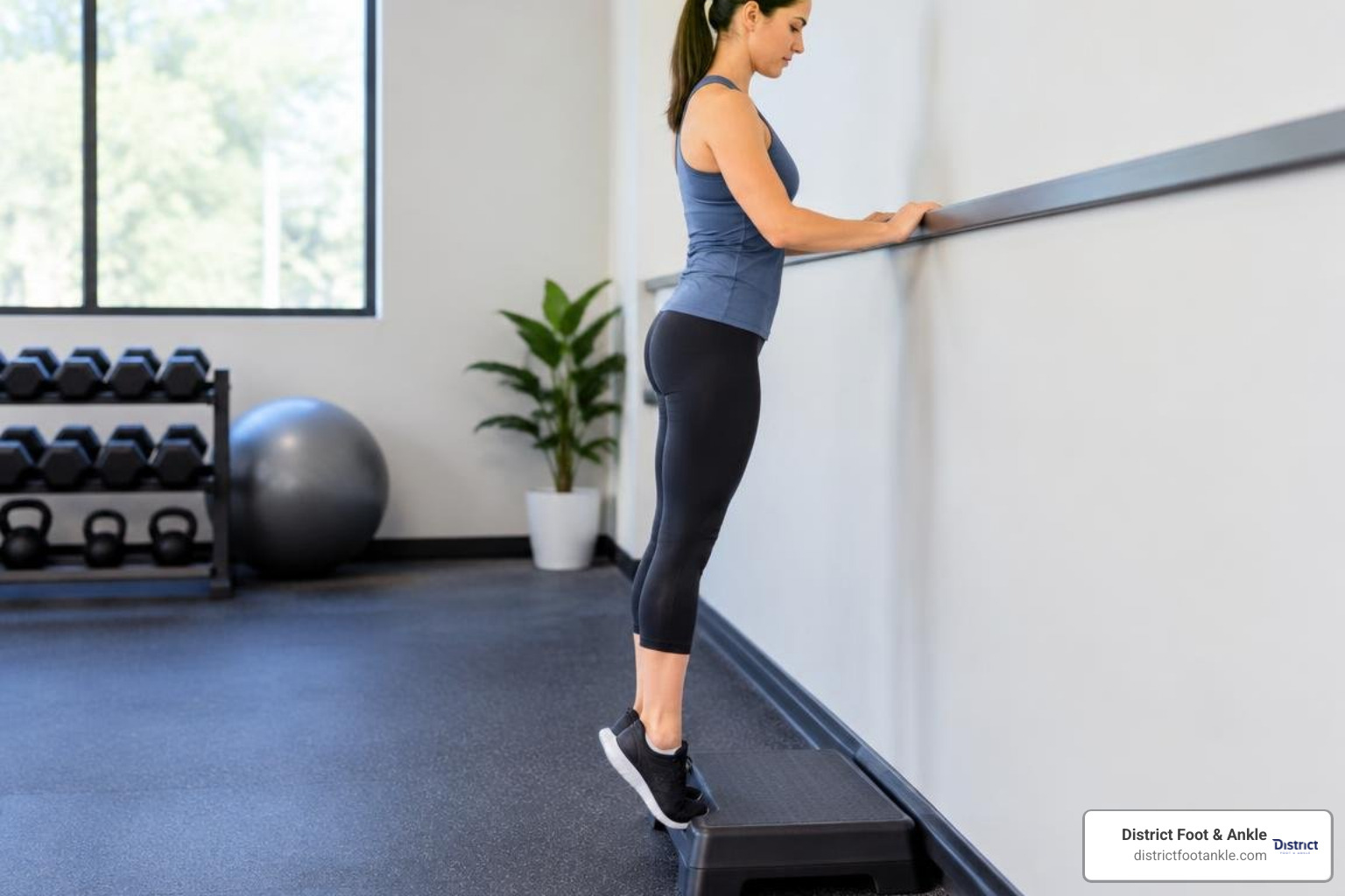
Effective rehabilitation of the gastrocnemius demands a structured transition from acute symptom management to progressive, high-load remodeling.
Acute Management (First 24–72 Hours)
For acute strains, the classic RICE protocol (Rest, Ice, Compression, Elevation) helps limit intramuscular hematoma expansion and excessive edema. Crutches or a temporary heel lift may be utilized to prevent excessive tension on the torn fibers.
Progressive Strength and Loading Continuum
Once acute pain subsides, therapeutic rehabilitation must systematically restore tensile strength, elasticity, and muscular endurance:
- Straight-Leg Isometrics: Seated or standing straight-leg isometric calf presses against an immovable resistance to stimulate cellular collagen synthesis without painful movement.
- Heavy-Slow Resistance (HSR) Eccentric Heel Drops: Performed with the knee completely straight to isolate the gastrocnemius (e.g., the Alfredson protocol). Lowering the heel below step level under control produces eccentric remodeling of the muscle-tendon fibers.
- Seated Bent-Knee Calf Raises: Performed with the knee bent at 90° to emphasize the deeper soleus muscle, ensuring balanced development across the entire triceps surae complex.
- Plyometric & Stretch-Shortening Drills: Incorporating jump roping, pogo hops, and bounding drills to restore elastic recoil and rate of force development (RFD).
- Clinical Modalities: Dry needling, instrument-assisted soft tissue mobilization (IASTM), and therapeutic ultrasound can help reduce chronic myofascial trigger points and improve soft-tissue glide.
Frequently Asked Questions About the Gastrocnemius Muscle
What is the primary difference between the gastrocnemius and soleus muscles?
The primary difference lies in their anatomical path and fiber composition. The gastrocnemius crosses two joints (knee and ankle) and contains predominantly fast-twitch (Type II) muscle fibers designed for high-velocity, explosive tasks like running and jumping. The soleus crosses only the ankle joint, lies deep to the gastrocnemius, and consists mainly of slow-twitch (Type I) fibers specialized for endurance and standing posture.
Does indoor cycling activate the medial gastrocnemius similarly to outdoor riding?
Yes. Electromyographic (EMG) studies demonstrate that medial gastrocnemius muscle activity is not significantly different between indoor stationary trainers and outdoor cycling (p = 0.246). In both settings, calf recruitment steadily climbs as perceived exertion (RPE) rises—ranging between ~24% and ~36% of maximum voluntary contraction.
How is a medial gastrocnemius strain diagnosed?
Diagnosis begins with a thorough physical exam assessing pinpoint tenderness along the medial calf belly, passive dorsiflexion pain, and weakness during single-leg heel raises. The clinician will conduct a Silfverskiöld test to check for contracture and a Thompson test to rule out an Achilles tendon rupture. Diagnostic ultrasound or MRI is frequently used to confirm tear severity (Grade I–III) and guide recovery timelines.
Conclusion
The gastrocnemius muscle is an architectural marvel of the lower leg, serving as a primary driver of human movement, athletic propulsion, and joint stability. Because it is subjected to high forces across both the knee and ankle, maintaining its flexibility, structural integrity, and tensile capacity is critical for athletes and active individuals alike.
If you are struggling with persistent calf pain, Achilles tightness, or limited ankle mobility, our team of board-certified podiatrists at District Foot & Ankle is here to help. Serving patients across Alexandria, Leesburg, and Northern Virginia, we combine advanced sports-medicine treatments and reconstructive expertise to create fast-track recovery plans. Schedule an appointment today for an expert evaluation of the gastrocnemius muscle and get back to moving pain-free.