

Could Gout Be Causing Your Swollen Ankle?
Yes. Gout can cause sudden ankle swelling, often with severe pain, heat, redness, and tenderness that makes shoes or even a bedsheet feel unbearable. Unlike a typical sprain, ankle gout may start overnight or without a clear twist or fall.
If your ankle becomes hot, red, rapidly swollen, and difficult to bear weight on, treat it as a medical concern. Rest the joint, elevate it, use a cold pack briefly, and contact a foot and ankle specialist promptly. Seek urgent care right away if you also have fever, chills, an open wound, spreading redness, or cannot move the joint, since infection can look similar to gout.
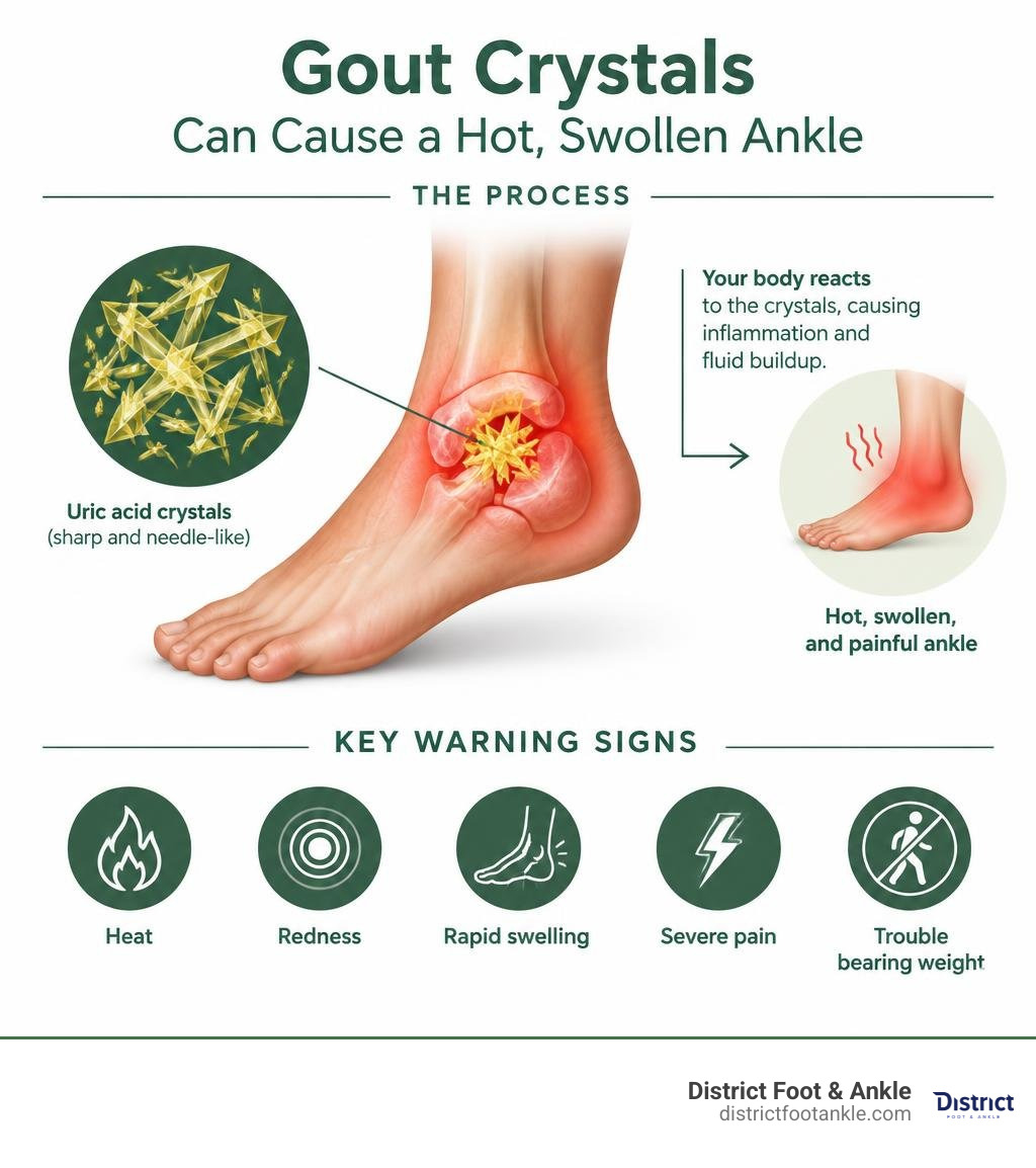
Gout happens when uric acid forms sharp crystals inside or around a joint. Your immune system reacts strongly to those crystals, causing inflammation and fluid buildup. While the big toe is the classic location, the ankle is involved in an estimated 12% to 15% of acute gout attacks.
I am Lonny Nodelman, DPM, FACFAS, a board-certified podiatrist with more than 13 years of experience treating foot and ankle conditions that limit movement. At District Foot & Ankle, I help active Northern Virginia patients sort out gout and ankle swelling from injuries and other causes, then build a practical path back to comfortable activity.
Understanding Gout and Ankle Swelling: Causes and Biological Mechanisms
To understand why gout targets the ankle, we have to look at the underlying biology. Gout is the most common inflammatory arthritis in adults, affecting an estimated 8.3 million people across the United States—roughly 4% of the adult population. Prevalence has nearly doubled over the past two decades, driven largely by shifting metabolic health and dietary trends.
The disease process begins with hyperuricemia, a state where serum uric acid levels exceed the normal solubility threshold of 6.8 mg/dL. When blood becomes oversaturated with uric acid, the excess precipitates out of solution as needle-shaped monosodium urate (MSU) crystals. These sharp crystals deposit directly into the synovial lining, cartilage, surrounding tendons, and soft tissues of your joints.
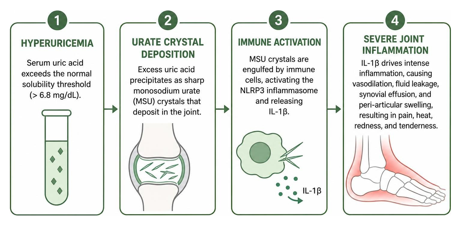
Once deposited, these crystals trigger an aggressive immune response:
- Inflammasome Activation: White blood cells (macrophages) engulf the microscopic crystals, which activates the intracellular NLRP3 inflammasome.
- Cytokine Cascade: This protein complex releases potent pro-inflammatory cytokines, primarily interleukin-1 beta (IL-1β).
- Synovial Effusion & Vascular Leakage: IL-1β triggers intense local vasodilation and vascular permeability. Plasma rushes into the joint capsule (creating an intra-articular synovial effusion) and leaks into the surrounding soft tissues (creating extensive peri-articular edema).
Why does this happen so readily in the ankle? Uric acid behaves much like sugar in a cup of coffee—it dissolves well in heat but rapidly turns gritty and precipitates when cooled. Peripheral extremities run cooler than the body’s core. The ankle joint typically sits at a physiological temperature between 32°C and 34°C (89.6°F to 93.2°F), significantly lower than core body temperature (37°C or 98.6°F). This localized cooling lowers the saturation point of uric acid, making the ankle an ideal environment for crystal precipitation.
Furthermore, the extensive soft tissue swelling seen in an acute flare presents as tense, non-pitting edema. Unlike systemic swelling from heart or kidney issues where pressing a finger leaves a prolonged indentation, gout-induced swelling is firm, taut, and hot to the touch. For more on the clinical mechanics of this presentation, explore scientific research on ankle gout manifestations.
How Gout and Ankle Swelling Develop Overnight
One of the most striking characteristics of an acute gout attack is how quickly it appears. Patients often go to bed feeling completely healthy, only to wake up around 2:00 a.m. or 3:00 a.m. with excruciating pain and an ankle that looks like an inflamed balloon.
Clinical data shows that the risk of an acute gout attack is roughly 2.4 times higher overnight and in the early morning hours compared to daytime periods. This nocturnal vulnerability stems from three primary physiological events:
- Peripheral Body Cooling: During deep sleep, ambient temperatures drop and the body redirects circulation inward, causing peripheral joint temperatures in the feet and ankles to dip even further.
- The Circadian Cortisol Nadir: Cortisol, your body’s natural anti-inflammatory hormone, drops to its lowest 24-hour concentration during the overnight hours, leaving inflammatory pathways unchecked.
- Relative Dehydration: Overnight fluid loss without fluid intake slightly concentrates serum urate levels, tipping marginal concentrations past the crystallization threshold.
As crystals precipitate within the joint space, the inflammatory surge causes rapid distension of the ankle capsule. Within just a few hours, the overlying skin becomes taut, shiny, and erythematous (bright red or purplish).
Common Triggers and Risk Factors in Men and Women
An acute flare in the ankle rarely happens without a catalyst. When hyperuricemia is present, specific triggers can suddenly destabilize urate levels and provoke crystal shedding:
- Dietary Purines: Consuming large quantities of purine-dense foods—such as organ meats, venison, red meat, mussels, scallops, and anchovies—causes a rapid spike in uric acid production.
- Alcohol Intake: Alcohol (especially beer, which contains high levels of brewer’s yeast and purines) is a dual threat. It stimulates uric acid production while dehydrating the body and impairing the kidneys’ ability to filter urate.
- High-Fructose Corn Syrup: Fructose metabolism in the liver rapidly consumes cellular ATP, directly generating uric acid as a byproduct.
- Dehydration & Fasting: Inadequate fluid intake or crash dieting spikes blood urate concentrations.
- Medications: Thiazide and loop diuretics (frequently prescribed for hypertension), low-dose aspirin, and niacin elevate urate levels by reducing renal clearance.
- Physical Trauma or Micro-Injuries: Even a minor bump, a strenuous run, or an awkward step can mechanically dislodge pre-existing, dormant urate crystals into the synovial space, setting off an acute inflammatory flare.
While men between ages 40 and 60 represent the majority of gout patients, women are equally vulnerable after menopause. Estrogen exerts a natural uricosuric effect, helping the kidneys excrete uric acid efficiently. Following menopause, estrogen levels drop, causing serum uric acid concentrations to rise toward male levels.
Because gout is still frequently mischaracterized as a “man’s disease,” women face substantial diagnostic hurdles. Studies reveal that women wait an average of 4.5 years longer than men to receive a confirmed gout diagnosis. This delay often leaves women managing painful, unexplained joint flares without targeted treatment.
Identifying Ankle Gout: Symptoms, Sensations, and Differential Diagnosis
When an acute gout flare strikes the ankle, it makes its presence known quickly. Patients frequently rate the pain between 8 and 10 on a standard visual analog scale. Common descriptions include feeling as though hot lava or crushed glass has been injected into the joint capsule.
The hallmark clinical signs of ankle gout include:
- Severe Allodynia: Extreme hypersensitivity where even a gentle breeze, a sock, or a light bedsheet resting on the skin triggers intense agony.
- Localized Heat and Erythema: The skin over the lateral and medial malleoli becomes bright red, shiny, and measurably hot (often 2°C to 4°C warmer than the unaffected leg).
- Significant Structural Distension: Rapid, non-pitting swelling obliterates the natural anatomical contours of the ankle bones.
- Profident Mechanical Restriction: Active and passive range of motion is severely limited by pain and fluid pressure within the joint.
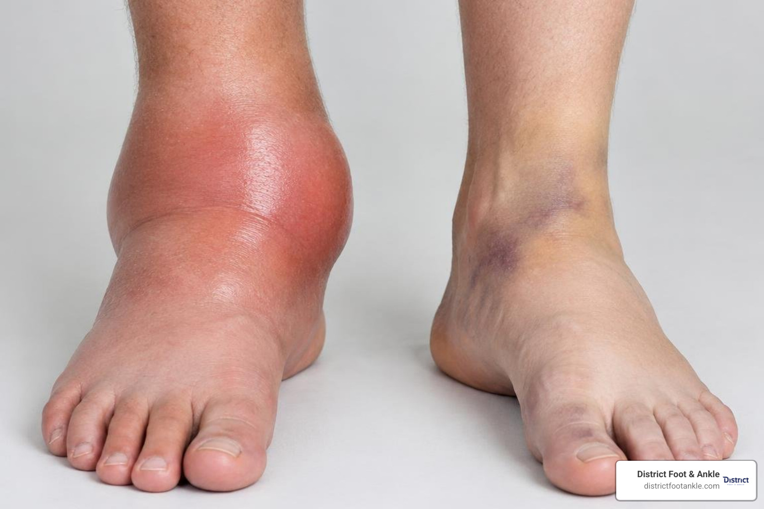
Because a hot, swollen ankle can mimic several other urgent conditions, an accurate differential diagnosis is critical. If you are experiencing unexplained joint swelling, reviewing our guide to foot and ankle pain relief can help you identify broader musculoskeletal symptoms.
Gout vs. Sprains, Cellulitis, and Septic Arthritis
Mistaking an ankle gout flare for a routine sprain or an infectious process is one of the most common diagnostic pitfalls in foot and ankle medicine.
| Diagnostic Feature | Acute Ankle Gout | Ankle Sprain | Cellulitis | Septic Arthritis (Medical Emergency) |
|---|---|---|---|---|
| Primary Cause | Monosodium urate crystal inflammation | Ligamentous tear/strain from trauma | Bacterial skin infection (Staph/Strep) | Bacterial infection inside joint space |
| Onset Speed | Explosive; peaks in 6–12 hours (often overnight) | Immediate at moment of trauma | Gradual; spreads over 24–72 hours | Rapid; worsens aggressively over hours |
| History of Trauma | Absent (or trivial micro-trauma) | Clear history of twisting, rolling, or impact | Absent (often follows a cut, scratch, or ulcer) | Absent (or recent joint injection/surgery) |
| Visual Appearance | Shiny, bright red or purplish; tense swelling | Ecchymosis (bruising), localized swelling | Diffuse spreading redness without sharp margins | Severe effusion, tight skin, extreme erythema |
| Local Temperature | Intensely hot to the touch | Mildly warm initially | Warm to hot across the affected skin | Burning hot over the entire joint capsule |
| Systemic Fever | Rare (occasional low-grade fever) | Completely absent | Common (mild to moderate fever) | High fever (>38.5°C/101.3°F), chills, and rigors |
| Pain with Light Touch | Extreme allodynia (bedsheet hurts) | Tender over specific ligaments on palpation | Tender to firm touch, but rarely allodynic | Extreme, incapacitating pain with any joint motion |
Attack Duration and Mobility Limitations
Left untreated, a standard acute gout flare in the ankle typically peaks within 12 to 24 hours and persists for 7 to 14 days before the body’s native anti-inflammatory mechanisms begin clearing the fluid. However, the absence of acute pain does not mean the joint has fully normalized. Lingering low-grade stiffness, mild peri-articular puffiness, and skin desquamation (peeling) can persist for several weeks.
During the height of an attack, attempting to walk on the affected leg is rarely possible. The sheer pressure of bearing weight forces the inflamed synovial membranes against the sharp urate crystals, creating unbearable mechanical pain. Patients typically require complete rest, crutches, or a walking boot during the first 48 to 72 hours.
Clinical Diagnosis and Evidence-Based Treatment Options
Accurately diagnosing gout in the ankle requires clinical experience and targeted diagnostic testing. It is a common misconception that a blood test alone confirms or rules out a gout flare. In reality, serum uric acid levels can appear completely normal or low during an acute attack, because inflammatory cytokines prompt the kidneys to excrete higher volumes of urate in the urine.
To establish a definitive diagnosis, podiatric physicians rely on several established clinical tools:
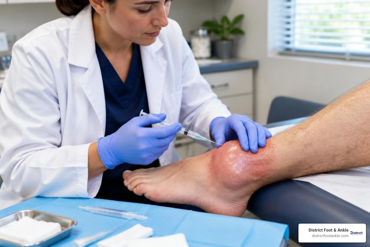
- Arthrocentesis (Joint Fluid Aspiration): The absolute gold standard. A small needle is inserted into the ankle joint to withdraw a sample of synovial fluid. Under polarized light microscopy, monosodium urate crystals appear as brightly glowing, needle-shaped crystals with negative birefringence (turning yellow when aligned parallel to the compensator axis).
- High-Resolution Musculoskeletal Ultrasound: A fast, non-invasive imaging tool that can detect the pathognomonic “double-contour sign”—an irregular, hyperechoic band of crystal deposition coating the surface of the articular cartilage.
- Dual-Energy CT (DECT): An advanced cross-sectional imaging method that uses two distinct X-ray spectra to map urate crystal deposits directly, color-coding them green inside the ankle joint without requiring a needle stick.
- Conventional Radiographs (X-rays): While standard X-rays do not show early crystal deposition, they help rule out acute fractures and evaluate for the classic “punched-out” erosions with overhanging bony edges characteristic of chronic tophaceous gout.
For additional clinical perspectives on identifying these symptoms early, review these clinical insights on ankle flare warning signs.
Immediate Relief and In-Clinic Interventions
When a patient arrives at our Alexandria or Leesburg clinics in the throes of an acute flare, our goal is to halt the inflammatory cascade quickly:
- Colchicine: Highly effective if taken within the first 12 to 24 hours of flare onset. It works by disrupting microtubule assembly in white blood cells, preventing them from attacking the urate crystals. Modern evidence-based dosing uses a low-dose protocol (1.2 mg at first sign, followed by 0.6 mg one hour later) to minimize gastrointestinal side effects.
- Prescription NSAIDs: High-dose anti-inflammatories such as indomethacin, naproxen, or celecoxib help suppress prostaglandin synthesis and relieve swelling.
- Corticosteroids: For patients who cannot take NSAIDs due to kidney disease, ulcers, or cardiovascular risks, a short course of oral prednisone provides potent, broad-spectrum anti-inflammatory relief.
- Intra-Articular Injections: In severe presentations where systemic medications are contraindicated, our specialists can perform an ultrasound-guided joint aspiration to relieve mechanical fluid pressure, followed immediately by an injection of a localized corticosteroid to calm inflammation at its source.
At-Home Remedies and Immediate Care Protocols
While coordinating medical evaluation, you can implement conservative steps at home to alleviate discomfort:
- Strict Elevation: Elevate the ankle on several pillows above heart level. This uses gravity to clear inflammatory interstitial fluid and reduce throbbing pressure.
- Cold Therapy: Apply a cold pack wrapped in a thin towel for 15 to 20 minutes every two hours. Never apply bare ice directly to the skin. Cold helps numb nerve endings and blunt inflammatory vasodilation.
- Aggressive Hydration: Drink at least 8 to 10 glasses of water daily. Ample hydration supports renal clearance of uric acid and helps prevent crystal formation in the kidneys.
- Protect the Joint from Contact: Use a wire basket, a firm pillow, or a tented cardboard box under your bedsheets to create a “bedsheet cradle,” keeping heavy linens from resting on the hypersensitive joint.
- Wear Wide, Open Footwear: Avoid constricting shoes or tightly laced sneakers. Switch to an adjustable slide or an open-toed recovery sandal until the swelling clears.
Long-Term Management to Prevent Gout and Ankle Swelling
Extinguishing an acute flare is only the first step. Preventing permanent joint damage requires Urate-Lowering Therapy (ULT) to lower systemic uric acid below the crystallization threshold:
- Target Serum Urate Levels: The clinical target for effective long-term disease control is a serum uric acid level consistently below 6.0 mg/dL (and below 5.0 mg/dL for patients with visible tophi or structural joint erosion). At these levels, new crystals cannot form, and existing crystals slowly dissolve out of the joint tissues over time.
- Allopurinol: The first-line xanthine oxidase inhibitor used globally. It works by blocking the biochemical pathway responsible for producing uric acid. Doses are carefully titrated upward while monitoring blood work.
- Febuxostat: An alternative xanthine oxidase inhibitor used for patients who cannot tolerate allopurinol or who require stronger urate suppression.
- Uricosuric Agents: Medications like probenecid that prompt the kidneys to filter and excrete higher volumes of uric acid.
Long-term management requires consistency. Stopping urate-lowering medication abruptly during an acute attack can destabilize blood urate levels and worsen the flare. Any changes to maintenance therapy should always be supervised by your doctor.
When to Seek Medical Attention and Emergency Warning Signs
Ignoring repeated gout flares in the ankle can lead to irreversible joint complications. Over months and years of chronic inflammation, urate deposits coalesce into hard, chalky masses known as tophi. Tophi can infiltrate the Achilles tendon, peroneal tendons, and the ankle joint capsule itself, acting like coarse sandpaper against delicate soft tissues.
Chronic, uncontrolled gout leads to:
- Severe Cartilage Destruction and Bone Erosion: Crystals erode the joint margins, causing permanent joint space narrowing and chronic secondary osteoarthritis.
- Tendon Rupture: Chronic tophaceous infiltration can weaken the Achilles or peroneal tendons, leading to spontaneous tearing or functional loss.
- Deformity and Chronic Instability: Progressive architectural damage can destabilize the ankle mortise, resulting in persistent gait abnormalities and chronic pain.
Critical Red Flags and Emergency Symptoms
While gout is painful, certain symptoms suggest a potentially life-threatening joint infection (septic arthritis) or spreading infection that requires immediate emergency evaluation.
Go to an emergency room or seek urgent medical attention immediately if you notice:
- A body temperature exceeding 38.5°C (101.3°F), accompanied by shaking chills or rigors.
- Rapidly spreading redness, red streaks tracking up the calf, or blister formation over the skin.
- Purulent (cloudy, thick, or yellowish) drainage oozing from the joint or skin.
- Complete inability to move the ankle joint even a fraction of an inch due to unbearable, locked agony.
- Severe swelling accompanied by sudden shortness of breath or chest pain (which could indicate a deep vein thrombosis or pulmonary embolism).
Frequently Asked Questions About Gout and Ankle Swelling
Can you walk with gout in your ankle?
During the acute phase of an attack, walking is rarely possible and generally ill-advised. Bearing weight compresses inflamed synovial tissues against sharp monosodium urate crystals, intensifying tissue trauma and worsening pain. We recommend resting the limb, elevating the foot, and using crutches or a knee scooter until the acute inflammation is controlled with medication.
How long does an ankle gout attack last if untreated?
Without medical intervention, an acute ankle flare typically lasts between 7 and 14 days. Pain usually peaks within the first 12 to 24 hours, followed by a gradual reduction in redness and heat. However, low-grade swelling, joint stiffness, and tissue tenderness can linger for several weeks. Prompt treatment with prescription anti-inflammatories or colchicine within 24 hours can significantly shorten the attack.
Can women get gout in the ankle?
Yes. While premenopausal women have lower rates of gout due to the protective uricosuric effects of estrogen, their risk rises significantly after menopause. Postmenopausal women make up a substantial portion of chronic gout cases. Because symptoms may be misattributed to osteoarthritis or a sprain, women experience an average diagnostic delay of 4.5 years. Any sudden, hot, unexplained ankle swelling in women should be evaluated for crystal arthritis.
Conclusion
An ankle that suddenly becomes hot, swollen, and exquisitely tender is not something to brush aside as a routine sprain. Left untreated, recurring crystal deposition can quietly degrade cartilage, erode bone, and damage surrounding tendons. Recognizing early warning signs and confirming an accurate diagnosis is the first step toward long-term joint health.
At District Foot & Ankle, our board-certified podiatric team provides comprehensive diagnostic assessments—including high-resolution imaging and in-office joint fluid analysis—to pinpoint the cause of your joint pain and create a clear, personalized treatment plan. Whether you are managing an acute flare or looking to prevent future joint damage, our clinics in Alexandria (4875 Eisenhower Avenue, Suite 230) and Leesburg (224D Cornwall St NW, #203) are here to help you get back to comfortable, pain-free movement.
Take control of your joint health today by identifying common causes of gout in the ankle and scheduling a focused clinical consultation with our foot and ankle specialists.