
You rolled your ankle on a curb two days ago and it is still swollen. Or the ache under your forefoot has been there for six weeks and nothing has explained it. Either way you are wondering the same thing: should somebody take a picture of this, and if so, which kind, and what is it going to cost?
Choosing between an X-ray or MRI for foot pain is not really a patient decision, but understanding how the choice gets made is useful, because it tells you what to expect at the visit and what to ask about before anything is ordered. Here is what each study shows, when imaging gets skipped entirely, and what the money side looks like.
Table of Contents
- Why you may not need imaging at all
- What a foot or ankle X-ray actually shows
- What an MRI shows that an X-ray cannot
- How the choice between an X-ray or MRI for foot pain gets made
- What imaging costs, and the question to ask before an MRI is ordered
- What to do if you were sent home without imaging and the pain is not settling
- Frequently Asked Questions About X-Ray and MRI for Foot Pain
- Getting seen in Alexandria and Leesburg
Why you may not need imaging at all
This surprises people, but most ankle injuries are not fractures. In the research behind the Ottawa ankle rules, a widely used clinical decision tool, the overall incidence of fracture in ankle injuries is under 15 percent. That is exactly why the rules were developed: clinicians were X-raying nearly everyone, and most of those images were normal.
The rules give a clinician a structured way to decide, based on where the pain is on the bone and whether you can put weight through the foot. They are a clinical tool used during an exam, not a self-test. Do not use them to talk yourself out of being seen, particularly if the pain is not settling or you cannot walk on it.
What this does mean is that arriving expecting an X-ray, and being told you do not need one, is a legitimate outcome rather than a brush-off. A careful history and a hands-on exam answer a lot of foot questions without a single image. If a twisted ankle is what brought you here, there is more on ankle sprains and how they are assessed on our ankle sprain page.
What a foot or ankle X-ray actually shows
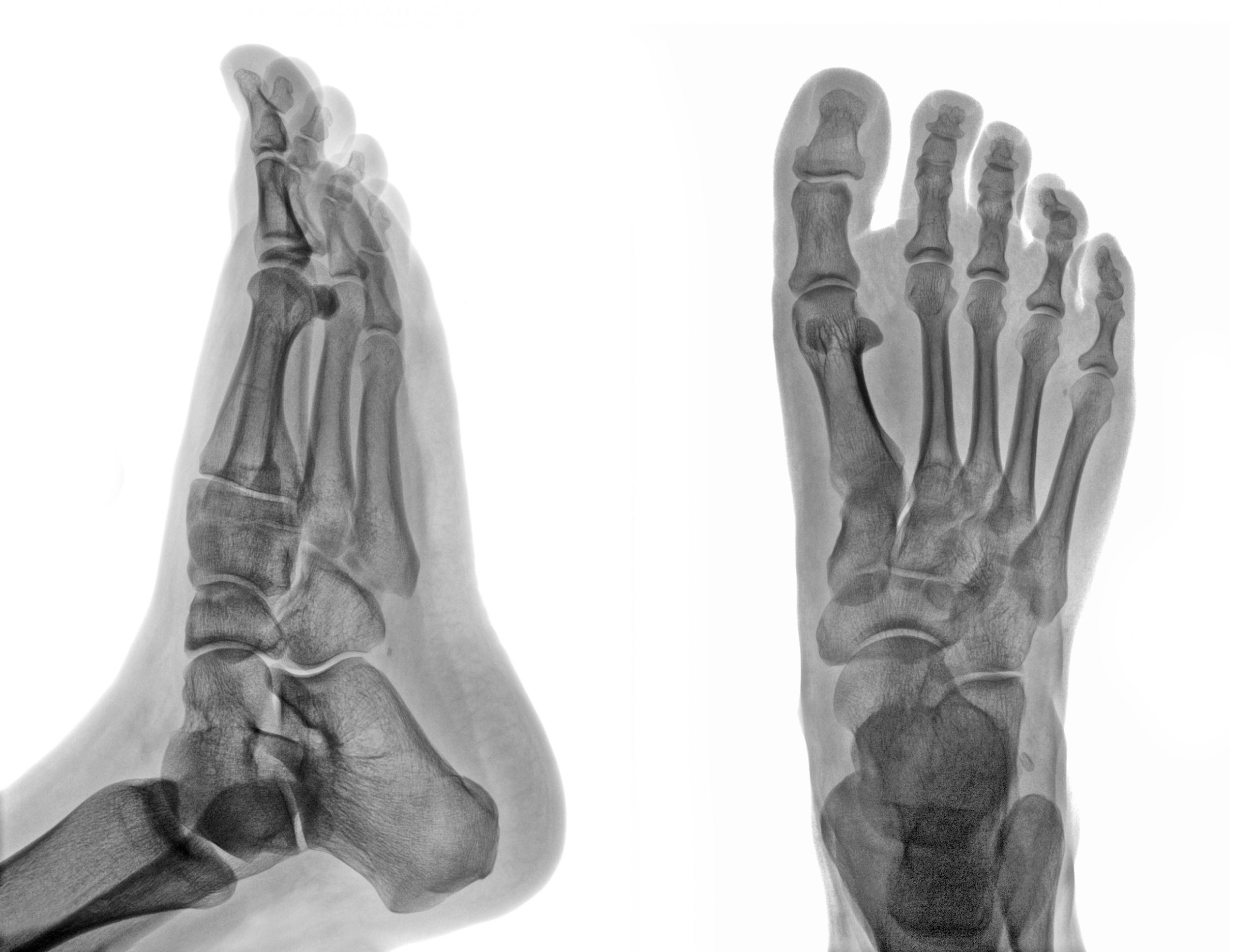
An X-ray is a bone picture. It shows fractures, dislocations, joint space narrowing from arthritis, bone spurs, the alignment of a bunion or a hammertoe, and hardware from previous surgery. It is fast, it is low cost, and it is usually the first study for a good reason.
It also has a specific and useful trick: weight-bearing views. Standing on the foot while the image is taken shows how the bones actually sit under load, which is how you live in your feet. A bunion or a collapsing arch can look very different standing than lying down, and for a deformity question the weight-bearing view is often the one that matters.
What an X-ray does not show well is soft tissue. Ligaments, tendons, the plantar fascia, cartilage and bone marrow are largely invisible on a plain film. A hairline stress fracture can also be invisible for the first couple of weeks, only becoming apparent once healing starts to show.
What an MRI shows that an X-ray cannot
An MRI images soft tissue and the inside of bone. It is the study that answers questions like: is this tendon torn or just inflamed, is there a stress reaction in this bone before it becomes a visible fracture, is the cartilage damaged inside this joint, is there a soft tissue mass, is there an infection tracking into bone.
It is slower, substantially more expensive, and it is not a better version of an X-ray. It is a different instrument answering different questions. For a straightforward acute injury where the question is “is this bone broken”, an MRI is usually the wrong first tool, not the premium one.
There are other studies in the mix too. CT is used for detailed bone architecture, particularly in complex fractures and pre-surgical planning. Diagnostic ultrasound has a role in looking at certain tendons and soft tissue in real time. Which studies are performed on site versus arranged elsewhere varies by practice, so it is a fair question to ask when you book. You can also see the services we provide in office before you call.
How the choice between an X-ray or MRI for foot pain gets made
Roughly, the sequence runs like this. The history and exam come first and generate a specific question. If that question is about bone, alignment, or an acute injury, a plain X-ray usually answers it. If the X-ray is normal but the clinical picture still points at something structural, or if the question was always about soft tissue, that is when advanced imaging enters the conversation.
The order matters for two practical reasons. One, the cheap study frequently answers the question, so starting there saves you money. Two, insurers generally expect that sequence, and an MRI requested without a preceding X-ray and a documented clinical rationale is far more likely to be delayed or denied.
If you are two days out from a twisted ankle and someone offers an MRI as a first step, it is reasonable to ask why.
Not sure whether what you are dealing with warrants a visit at all? Book an appointment at our Alexandria or Leesburg office and we can tell you after an exam, or call 703-832-9013 first.
What imaging costs, and the question to ask before an MRI is ordered
We post our self-pay prices, and we do that deliberately. A foot or ankle X-ray at three views is listed at 55.00 dollars, and a new patient examination at 300.00 to 450.00 dollars. As that page states, we are unable to determine final out of pocket costs prior to patient examination by a physician, and a physician may recommend services beyond the ones listed.
An MRI is a different conversation, and we are not going to quote you a number for one, because we do not set it. It is performed and billed by an imaging facility, the price varies enormously between a hospital outpatient department and a freestanding imaging centre, and what you owe depends on your plan design and your deductible status.
So here is the practical sequence. Before an MRI is ordered, ask three questions. Does this need prior authorization under my plan. Which facility are you sending me to, and is there a lower-cost in-network option. What is the facility’s self-pay or cash price, in case that is cheaper than running it through my deductible.
One thing worth knowing about that first question: approval is not payment. HealthCare.gov’s own definition of preauthorization says plainly that it is a decision about medical necessity and is not a promise your plan will cover the cost. People are caught by that distinction constantly, and it is a common source of the surprise imaging bill.
If any of that feels awkward to ask, it should not. Asking what something costs before it happens is the most ordinary thing in the world everywhere except healthcare, and a practice that cannot answer it, or that treats the question as rude, is telling you something.
What to do if you were sent home without imaging and the pain is not settling
Getting no imaging on day one is often correct. Getting no imaging on day twenty-one, when the pain has not improved, is a different situation.
A reasonable rule of thumb: if a foot or ankle problem is not clearly improving over two to three weeks of sensible self-care, or if it is getting worse at any point, that is a reason to be re-examined rather than to keep waiting. Stress fractures in particular tend to announce themselves late, and a normal early X-ray does not rule one out.
Come back sooner than that, or seek care the same day, if you cannot bear weight, if the foot or ankle looks deformed, if there is spreading redness or a fever, or if you have diabetes and there is any new sore, wound, or change in colour or sensation. Diabetic feet have a shorter safe waiting window than everyone else’s, because reduced sensation can mask how serious something is.
Frequently Asked Questions About X-Ray and MRI for Foot Pain
Do I need an X-ray for a sprained ankle?
Often not. Most ankle injuries are not fractures, and clinicians use a structured exam, including where the bone is tender and whether you can bear weight, to decide. If the exam suggests bone involvement, or if the pain is not settling over a couple of weeks, an X-ray becomes worthwhile. The decision is made at the visit, not before it.
What is the difference between an X-ray and an MRI for foot pain?
An X-ray images bone: fractures, arthritis, alignment, spurs, hardware. An MRI images soft tissue and the inside of bone: tendons, ligaments, cartilage, stress reactions, infection. They answer different questions rather than sitting on a quality scale. For a suspected broken bone, the X-ray is the right tool, not the cheap one.
Can an X-ray miss a fracture?
Yes. Hairline and stress fractures are frequently invisible on a plain film in the first one to two weeks, becoming visible only once healing changes the bone. That is why a normal early X-ray combined with pain that is not improving is a reason to be re-examined rather than reassurance that nothing is wrong.
Should I ask for an MRI?
You can always ask why a particular study is or is not being ordered, and a good clinician will explain it. Asking for an MRI first, before an exam and usually before a plain X-ray, tends to slow things down rather than speed them up, because insurers generally expect that sequence and documented rationale.
What does a foot X-ray cost?
Our posted self-pay price for a foot or ankle X-ray at three views is 55.00 dollars, alongside a new patient examination at 300.00 to 450.00 dollars. Final out of pocket costs cannot be determined before a physician examination, and if you are using insurance what you owe depends on your plan and deductible status.
Does my insurance have to approve an MRI first?
Many plans require prior authorization for an MRI. Ask your insurer directly, before the study is scheduled. Also worth knowing: approval and payment are not the same thing. HealthCare.gov’s own definition notes that preauthorization is a medical-necessity decision and is not a promise your plan will cover the cost.
Getting seen in Alexandria and Leesburg
Our Alexandria office at 4875 Eisenhower Avenue, Suite 230 sits in the Eisenhower Valley corridor, which for most of the Northern Virginia patients we see means the visit is a short detour off the Beltway or up Route 1 rather than a trip into the city. That is a practical consideration with an injury: the whole point of getting a foot looked at quickly is that it is quick, and a forty-minute each-way drive is how a two-day-old ankle becomes a two-week-old ankle.
Our Leesburg office at 224D Cornwall St NW, Suite 203 serves the Loudoun corridor on its own terms rather than as an overflow. Ashburn, Sterling, Lansdowne and Purcellville patients are not going to drive east for an X-ray, and they should not have to. Both offices run Monday through Friday, 8 AM to 4 PM, on one number: 703-832-9013.
Book an appointment at whichever office is closer, or call and ask what an X-ray would cost before you come in. We would rather have that conversation on the phone than have you skip the visit over a number nobody would tell you.
Medically reviewed by Dr. Sammar Abueldoleh, DPM on September 28, 2026. Dr. Abueldoleh is Board Qualified in Foot and Ankle Surgery, with residency training at Legacy Health and Kaiser Northwest Permanente.
Not medical advice. This article is general education and does not diagnose any condition or recommend imaging for any individual. Talk with a qualified clinician about your own situation.