
Heel pain is one of the most common reasons why patients meet with Dr. Nodelman at District Foot and Ankle.
There are many causes of heel pain. In a another blog article, I list 5 other reasons why a patient might have heel pain that is not due to plantar fasciitis.
Plantar fasciitis does remain the most common cause of heel pain. This is characterized by pain in the bottom of the heel. It is typically exacerbated with first step in the morning but can improve. Depending on level of activity throughout the day, pain may worsen with time.
If symptoms have been going on for some time, it is important to obtain some imaging of the foot and/or ankle. The first place to start would be conventional radiographs of the foot. Radiographs are most important for ruling out certain issues like a stress fracture of the heel bone or a tumor.
Regular x-rays are not particularly useful for assessing the plantar fascia because x-rays do not show soft tissue structures.
An ultrasound is a very convenient way to assess soft tissue structures in the foot and the ankle. This also includes the plantar fascia. As plantar fasciitis becomes more chronic, the plantar fascia will demonstrate characteristic changes on an ultrasound including thickening and areas of degeneration. This is usually not seen in the more acute phases of plantar fasciitis.
The plantar fascia is normally less than 4 mm in thickness.
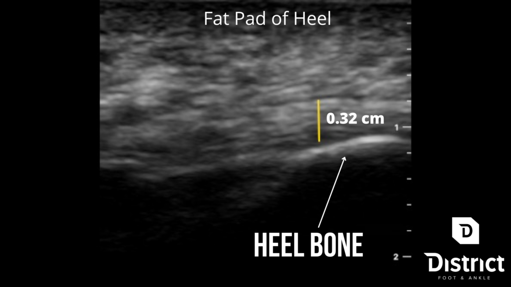
Here is an example of a normal fascia (this is Dr. Nodelman’s foot):
The image is upside down so the bottom of the foot is at the top part of the image. As you progress deeper, you reach the fat pad of the heel and eventually you reach the plantar fascia which is marked by the yellow line. In this case, Dr. Nodelman’s plantar fascia measures 0.32 cm which is a normal thickness (he also doesn’t not have plantar fasciitis). Deeper to the plantar fascia is the heel bone which is the horizontal white structure. The heel bone is also known as the calcaneus in medical terms.
Here is an example of a diseased plantar fascia:
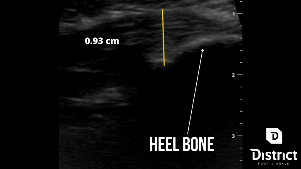
The other thing that can be noticed between the normal and abnormal fascia is that there are more areas of “black” in the thicker, disease plantar fascia. In radiological terms, this is referred to as increased hypoechogenicity and typically implies increased degeneration and abnormality of the soft tissue.
As you can see, ultrasound is more useful than an x-ray at assessing the plantar fascia in the setting of plantar fasciitis. X-rays are important, but they do not help with assessment of the plantar fascia, because as stated above, soft tissue structures do not show up on a conventional x-ray.
If a patient is experiencing significant heel pain, it is important to take an extensive history and perform a focused and detailed physical examination. To complete the picture, imaging should be performed. If the patient presents with what was previously diagnosed as plantar fasciitis, but there is no thickening or other characteristic changes on ultrasound that you would normally see with fasciitis, other diagnoses should be strongly considered.
As a teaser, I do have plans in the future to submit a post about how the “dreaded heel spur” is realistically of no clinical significance. Topic for another day!